


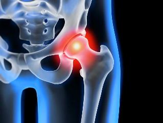
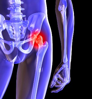
Hip osteoarthritis — degenerative-degenerative pathology, which is characteristic of hyaline cartilage itself refraction. The disease develops slowly, the accompanying pain syndrome and decreased range of motion. After a few years of absence of osteoarthritis in the initial stages of a medical intervention's thigh atrophy Nov. Limb shortened and the mixing of the damaged joint, which causes partial or complete paralysis of the hip joint. Into pathology reasons previous injury, curvature of the spine, the system Nov-diseases of the skeletal system.
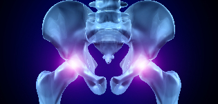
Patients with osteoarthritis usually taped middle-aged and elderly people. The results of instrumental diagnosis on the basis of exposure to research — radiography, MRI, computed tomography, computed tomography systems. 1 and 2 conservative treatment pathology severity. When detected, ankylosis, or lack of pharmaceutical treatment etc surgery (arthrodesis, arthroplasty).
Pathology development mechanism
Two bones that make up the hip joint — the first and thighs. Sub-section engages the iliac bone body shaping networks offered with the thighs, the upper portion of the acetabulum breakdown. Fovea of the femoral head during movement and the Joint still moves freely. This "returns" the device is flexible to allow for the hip joint, stretch, and encourage you to bring abstraction to return to hip. Slip joint structures that provides seamless, soft, flexible yellow elastic cartilage, which I started, and the femur bone. Its basic function re-loads the fast wear of the bone tissue alert while driving.
Under the influence of internal and external factors nutrients from the broken cartilage. No special circulatory system — synovial fluid: provides food texture. Osteoarthritis that becomes viscous thickening. Nutrient deficiency provokes a drying surface resulting in hyaline cartilage. In a closed fracture, resulting in a continuous micro-tissue injury of the hip joint in flexion or extension. Cartilage thinning, believe in tomorrow, its cushioning features. To "Harmony" the increasing pressure of the deformed bone. In the background, and degradation, destructive and degenerative changes progress in metabolic tissues.
Causes and precipitating factors
Primary or idiopathic osteoarthritis from developing without any reason. Accepted, destruction of cartilage tissue of the body slowing the aging process is the result of natural recovery in order to reduce the production of collagen and other compounds required for a complete regeneration of the hip joint structures. Secondary osteoarthritis on a background of a pathological situation is caused by already present in the body. Secondary diseases of the most common reasons include:
- previous injury — damage to the tendon apparatus, torn, Nov, their full breakup of the base of the bone fractures, dislocations;
- developmental articulation disorder, congenital dysplastic disorders;
- autoimmune pathology of rheumatoid jet, psoriatic arthritis, systemic lupus erythematosus;
- nonspecific inflammatory diseases such as pyogenic arthritis;
- specific infections — gonorrhea, syphilis, brucellosis, ureaplasmosis, Trichomonas, tuberculosis, osteomyelitis, encephalitis;
- violation of the functioning of the endocrine system;
- degenerative-degenerative pathology — osteochondropathy of the femoral head;
- hypermobility of joints on the production due to "stress" the collagen, thought-provoking their hyperactivity, fatigue bond.
Because it can cause for the development of osteoarthritis hemarthrosis (bleeding into the hip joint cavity), provocative factors, including blood disorders. This formation diseases, excessive weight, excessive exercise, sedentary lifestyle. Its development lead to the wrong organization, sport, lack of exercise, foods with a high content of dietary products, giraud, and water-soluble vitamins. Postoperative osteoarthritis is the result of, a few years later, after the surgery, especially bulky fabrics used accompanied. Gave osteoarthritis in the hip joint can not be transmitted. But of course some of the innate characteristics (such as metabolic disorder, skeletal structure) significantly increases the possibility of its development.
Symptoms
Leading symptoms of osteoarthritis in the hip joint pain while walking in hip and knee joint. A person's rigidity, movements, stiffness, especially in the morning. Vary limp walk it begins to stabilize the joint of the patient. Over time, atrophy and deformation of the joint in such a way that the leg is shortened Nov. Another characteristic pathology — limitation unipolar hip. For example, when difficulties arise to sit on a stool, legs spread wide to the side.
Even "working" at home that osteoarthritis can be cured! Just remember, smear it once a day...
For the initial severity of osteoarthritis, characterized by recurrent pain after intense physical exertion with nature. Articulation a localized area and disappears after a long vacation they.
Osteoarthritis second-degree connection increases the intensity of the hip pain. Uncomfortable feelings arise, and even resting spread thighs and groin, reinforced lifting or increased motor activity. To resolve the hip pain, that starts man, but loose. Notes limit joint movement, especially internal rotation of the hip and lead.
The third degree of osteoarthritis, characterized by long-lasting and powerful, it is a pain. The difficulties that arise during the transaction, for this reason, a person have to use a walking cane or crutches. The front plane Offset is happening because of weakness of the pelvic bone outlet thigh Nov. To compensate occurring shortening, the patient leans over the feet while moving the damaged limb. This provokes a strong center of gravity Offset and increased joint loads. This stage is joint ankylosis osteoarthritis that develops.
| Degrees | Radiographic signs |
| First | Expression changes not significantly. Joint cleft moderate, irregular and incomplete destruction of the surface of the femur using narrowed. Internal or external bone growth in the acetabulum in the breakdown of small plan |
| Second | The joint space is significantly reduced because of irregular height may be considered. Bone head, slipped hips down, deformed, increase, its contours become jagged. Glenoid fossa bone growth on the surface of the inner and outer edges that are created |
| Third | Observed full or partial mixing of the joint. Of the femoral head has been greatly expanded. The entire surface breakdown of the acetabulum, more than one in bone growth |
Diagnosis
When placing the doctor considers the diagnosis, clinical findings, pathology, medical history, results of the external audit, patient and instrumental studies. The most informative radiography. With his help the hip joint installed in his course a scene assessed the situation, the damage degree of cartilage tissue, and in some cases, and the development is the reason. If virtualnoje curved groove and a flattened nerve tissue and the site CCD is increased, then most likely congenital dysplastic articular changes acceptable. Specifies a secure pertus caps hip disease. Vivi radiography provides post-traumatic osteoarthritis, despite the lack of story, previous illness, injury. Other diagnostic methods are also used:
- SA scans of osteophyte growth in the mouth helps to detect bone plates;
- MR held to assess the status of their degree, including connective tissue structures and pathological process.
If necessary, the inner surface s with arthroscopic inspection tool. An exception to the differential diagnosis of gonarthrosis , degenerative disc disease, lumbosacral or thoracic. Violation of radicular pain syndrome due to clinical findings of osteoarthritis came in under the camouflage, or inflammation of the nerve. Often a series of tests failed except neurogenic pathology. Necessarily the hip joint the hip joint bursitis differentiated upper osteoarthritis, ankylosing spondylitis, reactive arthritis. Biochemical studies of blood and synovial fluid held exceptions for autoimmune pathologies.
Surgical intervention
We will focus on the ineffectiveness of conservative treatment of a complex pathology County the processing is performed. Re-osteoarthritis articular cartilage, damaged tissue, without surgery, and dentures is not possible, but can be treated with the right approach, of course, all the right lifestyle, by medical prescription, therapeutic exercises in the classroom, regular courses ankara, intake, vitamins and proper nutrition of cartilage and stop the process of defeat and destruction of the hip joints.